


Dry Eye Services


We are the Western Slope leaders in Dry Eye care!
Read more ...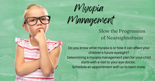
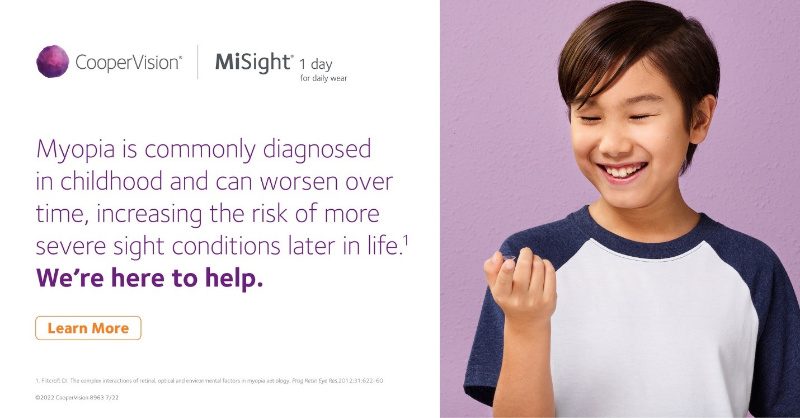
Myopia Management at Eyecare Specialties
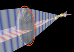
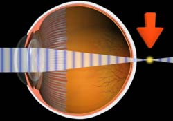
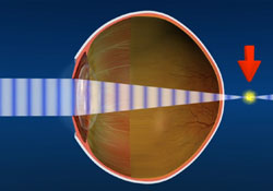
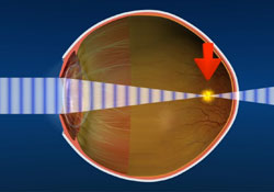
What is myopia?
Myopia is blurry long-distance vision, often called ‘short-sightedness’ or ‘near-sightedness’. A person with myopia can typically see clearly up close – when reading a book or looking at a laptop screen – but words and objects look fuzzy on a whiteboard, on television, across the room, when looking outdoors or when driving.
Why is myopia a concern?
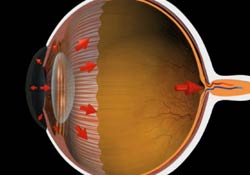
The rate of myopia is growing across the world. Most myopia is caused by the eye length growing too quickly in childhood. The eyes are meant to grow from birth until the early teens and then cease, but in myopia the eyes grow too much and/or continue growing into the teenage years. Once a child becomes myopic, their vision typically deteriorates every 6-12 months, requiring a stronger and stronger prescription. Most myopic children tend to stabilize by the late teens and early 20’s. Excessive eye growth raises concern because even small amounts of stretching can lead to increased likelihood of vision threatening eye diseases in later life, such as myopic macular degeneration, retinal detachment, and cataract.
Why manage myopia in children?
Myopia progresses fastest in younger children, especially those under age 10. This means that the most important opportunity to slow eye growth is when children are younger. Myopia management aims to apply specific treatments to slow the excessive eye growth to a lesser rate.
The short-term benefit of slowing myopia progression is that a child’s prescription will change less quickly, giving them clearer vision for longer between eye examinations. The long-term benefit is reducing the lifetime risk of eye disease and vision impairment
Treatments for slowing myopia progression
Standard, single-focus long distance spectacles or contact lenses do not slow down the progression of childhood myopia. Instead, specific types of spectacles, contact lenses and eye drops called atropine have been proven to slow myopia progression in children.
The best option for your child will depend on their current prescription and other vision and eye health factors determined in their eye examination. Your eye care practitioner will discuss the options with you to determine the best option.
Contact lenses
Standard single-focus contact lenses do not slow the worsening of childhood myopia but specific designs do. These specific designs can both correct the blurred vision of myopia and work to slow down myopia progression. The options include soft myopia controlling contact lenses and orthokeratology.
Orthokeratology
Orthokeratology (Ortho-k) is a non-surgical treatment in which you wear specially fitted contact lenses overnight while you sleep, to gently and safely reshape the front of your eyes. The lenses slowly reshape the cornea while you sleep, and in the morning, the lenses are removed. Your cornea will temporarily retain the shape of the lens, leaving you with a temporary correction that allows you to see clearly without glasses or contact lenses. Ortho-k is a popular treatment option, as it not only temporarily corrects myopia in the short-term, but can help reduce the lengthening of the eyeball with regular use.
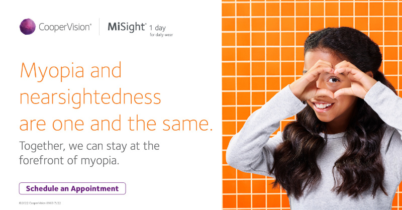
Multifocal Contact Lenses
Multifocal, soft lenses are special contact lenses that are designed with different powers in different areas of the lenses. MiSight 1 day® contact lenses are the only FDA approved soft contact lens indicated to slow the progression of myopia (nearsightedness) in children, aged 8-12 at the initiation of treatment. They are worn during waking hours. They are daily disposable. With daily disposable contact lenses, patients can experience clear vision, freedom from glasses and continue to enjoy the activities they love.
Atropine eye drops
Atropine eye drops in strong concentrations are used to temporarily dilate the pupil of the eye and stop the focussing muscles working in a variety of clinical applications. Atropine eye drops for myopia control, though, are a low-concentration with much fewer such side effects. Spectacles or contact lenses are still needed to correct the blurred vision from myopia, as atropine only acts to slow myopia progression.

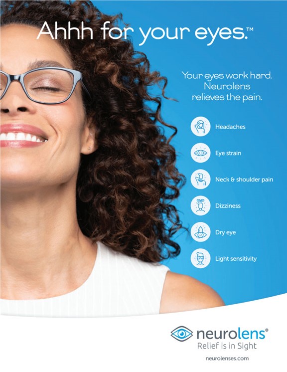
If you’re suffering from eye strain, headaches or neck pain, you’re not alone. You may be surprised to learn that about two out of three people experience symptoms of eye misalignment. The recent shift to remote and hybrid work has caused that number to increase, but many people don’t even know what eye misalignment is. Even minor misalignments can cause a variety of uncomfortable, painful symptoms that affect how you live and work.
The good news?
Relief is in sight and Neurolenses can truly change your life.
Do you suffer from any of these symptoms?
- Dry Eyes
- Eye Fatigue
- Eye Strain
- Headaches
- Neck Tension
- Shoulder Pain
- Motion Sickness
If so, you may be a good candidate for Neurolenses!
What Are Neurolenses? Eye misalignment occurs when your eyes and brain have difficulty working together to produce a single, clear image. This irritates the trigeminal nerve, which is attached to your brain and is the largest of the cranial nerves in your head. When your eyes are not properly aligned, additional pressure is put on the trigeminal nerve, leading to overstimulation and a range of uncomfortable symptoms. This is known as trigeminal dysphoria. Neurolenses are designed to reduce that pressure and bring your eyes into alignment using “contoured prism” technology. As more and more Americans begin to use computers, laptops and smartphones regularly, the number of people who experience symptoms of eye misalignment continues to grow.
Did you know that eye misalignment is actually a common underlying issue behind digital vision syndrome? If you frequently use digital devices, you’re probably familiar with symptoms such as dry eye, headaches, blurry vision and eye strain. The contoured prism design of Neurolenses brings your eyes into alignment and alleviates the symptoms that are holding you back from doing what you love. In fact, 93% of patients who have tried Neurolenses responded positively and said they experienced dramatic symptom relief within a matter of months!
How Does It Work?
 It all starts by taking the short, simple Neurolens Test to find out if you’re a candidate. Most people are, even if you don’t currently wear prescription glasses. Simply schedule an appointment with our team, and your eye care provider at Eyecare Specialties will be able to evaluate your symptoms and use the Neurolens Measurement Device to detect eye misalignment. This painless test only takes a few minutes to complete and makes it simple for your doctor to diagnose eye misalignment and get you well on your way to comfortable vision. Neurolenses are worn like your typical eyeglasses, and lenses can be customized to contain your current corrective prescription along with the correction needed to treat misalignment. They can also fit into most types of frames. If you don’t currently wear prescription eyeglasses, don’t worry! Neurolenses can still reduce the amount of pressure on your trigeminal nerve and help alleviate your symptoms
Let’s Get Started!
Are you ready to take the first step toward clear, comfortable vision? We’re here to help! At Eyecare Specialties, we’re dedicated to showing our patients results and improving lives. Give us a call at (970) 824-3488 or request an appointment online today. We look forward to seeing you soon!
Craig location's unique application URL:https://apply.sunbit.com/EyecareSpecialties-Craig
Steamboat Springs unique application URL:https://apply.sunbit.com/EyecareSpecialties-SteamboatSprings
Our knowledgeable staff at Eyecare Specialties will provide all paperwork necessary for you to seamlessly submit an out of network claim to your vision discount plan and any reimbursements available would be paid directly to you!
We strive to ensure that you understand your insurance coverage and will answer any questions you may have regarding your benefits.
Please contact our office to find out if your insurance provider is accepted in our office.
FSA/HSA
Many employers are offering Flexible Spending Account (FSA) or Health Savings Account (HSA) to employees. These plans are designed to let you save money in an account, pre-tax, to pay for additional medical expenses such as eye exams, glasses, contacts, and often laser vision surgery. Check with the benefits administrator at your work to see if you are eligible for this program.
Eyecare Specialties accepts CareCredit and Sunbit. CareCredit is a healthcare credit card with special financing options for healthcare purchases to help give our patients an additional way to pay for:
- Vision exams and more
- Multiple pairs of glasses
- Annual supplies of contact lenses
- Prescription and Plano sunglasses
- Corneal Refractive Therapy
- Designer Frames and lenses
- Dry Eye Services
We are the western slope leaders in Dry Eye care!
 At Eyecare Specialties we are committed to healthy vision. And as Dry Eye experts we offer specialized attention through our Dry Eye clinic.
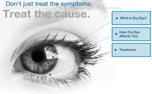
We understand your frustration with the limitations of traditional therapies and are committed to bringing you the most advanced technology available to treat this common disease – so that you can enjoy the greatest possible relief.
If you’ve been suffering with this painful condition – or think you may have it – we can determine the cause of your symptoms and apply therapy appropriate to your specific needs that may make your daily discomfort a thing of the past.
You Don’t Have to Live With Dry Eye Anymore
Dry, irritated eyes can be managed with new treatment that brings comfort and can restore quality of life.
Visit Eyecare Specialties to get an accurate diagnosis and a treatment plan tailored to your needs.
Dry eye is a long-term chronic disease that takes years to develop- and may worsen if left untreated.
There are two main forms of the disease: evaporative and aqueous deficient.
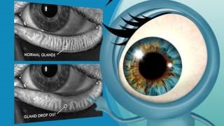
Evaporative Dry Eye
Evaporative Dry Eye is caused by blockages in the Meibomian glands located in your eyelids.
This condition of obstructed glands is known as Meibomian Gland Dysfunction (MGD). These glands are responsible for creating the lipid (oil) layer of tears. When the glands aren’t working as they should, you don’t have enough tear film oil.
Your tears- which lubricate your eyes and keep them comfortable-evaporate too quickly. An insufficient oil layer can cause your tears to evaporate 4-16 times faster.
Aqueous Deficient Dry Eye
Aqueous Deficient Dry Eye occurs when the lacrimal glands do not create a sufficient amount of aqueous (water) to keep the eyes moist.
Inadequate amount of tears. Tears are produced by several glands in and around the eyelids. Tear production tends to diminish with age or as a side effect of certain medicines. Environmental conditions, such as wind and dry climates, can also decrease tear volume due to increased evaporation. When the normal amount of tear production decreases or tears evaporate too quickly from the eyes, symptoms of dry eye can develop.
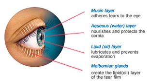 Poor quality of tears. Tears are made up of three layers: oil, water, and mucus. Each component protects and nourishes the front surface of the eye. A smooth oil layer helps prevent evaporation of the water layer, while the mucin layer spreads the tears evenly over the surface of the eye. If the tears evaporate too quickly or do not spread evenly over the cornea due to deficiencies with any of the three tear layers, dry eye symptoms can develop.
If you suffer from any of these symptoms from time to time....
you may benefit from a visit to our Dry Eye clinic.
- Dryness
- Discomfort and irritation
- Grittiness or feeling of a foreign body in the eye
- Burning or stinging sensation
- Tearing
- Redness
- Discharge
- Tiredness
- Itching
- Vision Disturbance
- Sensitivity to light
How do we determine if you have Dry Eye?
 In our Dry Eye Clinic, Dr. Eckroth will conduct an ocular surface disease evaluation and determine the likely causes of your dry eye symptoms. LipiView is a non-invasive, can be performed in our office, and takes less than 5 minutes. Lipiview takes an extremely detailed image of your eye’s tear film.
Calculations and images are instantly displayed on the LipiView monitor.
How Dry Eye can negatively impact your vision – and your life
The symptoms of Dry Eye can be uncomfortable – and a big burden.
Basic visual tasks, such as reading, using a computer, driving or watching television may become difficult. Wearing contact lenses may be impossible. And, you might find that symptoms worsen later in the day, keeping you from enjoying the activities you want to do.
Underlying the considerable discomfort is a real physical condition that needs treatment to stop the cycle of Dry Eye deterioration and worsening symptoms. Dry Eye is a chronic disease – and without proper management, the deterioration may look like this:
- Increased evaporation of tears
- Unstable tear film
- Damage to the eye surface
- Further discomfort
- Inflammation and cell damage
- Fluctuation and decrease of vision.
We have a variety of treatments available for you here at our Dry Eye Clinic.
Dry eye disease is a widespread and common issue for up to 49 million Americans. This chronic condition can have a significant impact on your quality of life, causing a foreign body sensation in your eyes, pain, blurry vision, and dry or watery eyes. Untreated, it can even lead to further eye health complications.
Despite these constant detrimental effects on quality of life, many dry eye sufferers are not aware that they’re suffering from dry eye disease or that real treatments exist. Instead, they just live with the discomfort.
At Eyecare Specialties, we have a team dedicated to improving your quality of life. If you suffer from dry eyes, we can determine the cause of your problems using the latest technology available. We can recommend a customized treatment plan designed to relieve your dry eye symptoms.
- BLEFEX : The first and Only Doctor Eyelid Cleaning Procedure to help maintain clean and healthy lids.
- LIPIFLOW THERMAL PULSATION SYSTEM : is a significant technological shift managing evaporative dry eye. Applying a combination of directed heat and pulsatile pressure, many find this effective in relieving the blockage of their meibomian glands.
- INTENSE PULSED LIGHT (IPL) treatment is lightly applied to the skin to treat inflammation by stimulating the body’s natural healing process.
BlephEx®
The first and only doctor eyelid cleaning procedure to help maintain clean and healthy lids.
BlephEx® uses a soft rotating medical grade micro-sponge along the edge of your eyelids and lashes. This procedure removes excess bacteria, biofilm and bacterial toxins, which are the main causes of eyelid symptoms.
LipiFlow Thermal Pulsation System®
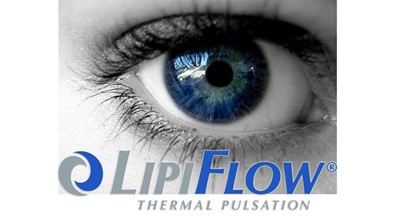 If you’ve been unable to receive long-lasting relief from over-the-counter or prescription remedies you’ve tried, it may be because they haven’t been targeting the primary cause of your condition – the blockage of your Meibomian glands located in your upper and lower eyelids. Perhaps you have tried things like eye drops and warm compresses, which many find inconvenient and only offer short-term relief.
LipiFlow® is a treatment device that applies heat and massage to both the outer and inner eyelids. This application has proved to be an extremely effective process for clearing blockages found in the Meibomian glands, allowing them to produce the oils and function properly.
What Is the OptiLight by Lumenis & How Can It Manage Your Dry Eye?
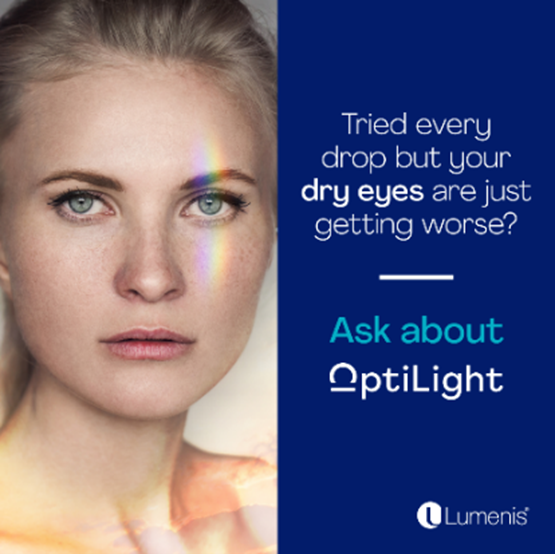 We’re proud to offer OptiLight by Lumenis to our patients, specially designed for dry eye management.
OptiLight by Lumenis is a light-based, non-invasive treatment done in the area below the eyes to manage dry eye. The first and only IPL FDA-approved for dry eye management.
The treatment is safe, gentle, and is backed by more than 20 clinical studies.
How Does It Work?
OptiLight uses precise pulses of light to reduce the inflammation that is typically associated with dry eye disease, improve tear break-up time, and increase meibomian gland functionality.
This application can significantly relieve dry eye indicators and has a multi-factorial effect, including:
- Increasing tear break-up time
- Reducing the amount of demodex mites and bacteria living around your eyes
- Eliminating blood vessels that contribute to inflammation
- Improving meibomian gland functionality
 In our continued efforts to bring the most advanced technology available to our patients, Drs Eckroth and Vides recommend the Optomap as an integral part of your eye exam.
Many eye problems can develop without warning and progress with no symptoms. Early on, you might not notice any change in your vision. However, diseases such as macular degeneration, glaucoma, retinal tears or detachments, as well as other health problems such as diabetes and high blood pressure, can often be detected with a thorough exam of the retina. The retina is the part of your eye that catches the image of what you are looking at, similar to the film in a camera.
An Optomap provides:
- A 200 degree laser, digital, image of the retina to confirm a healthy eye or detect the presence of disease.
- A map of the retina, giving your eye doctor a more detailed view than he can achieve by other means.
- The opportunity for you to view and discuss the Digital images of your eye with your doctor at the time of your exam.
- A permanent record for your medical file, enabling your optometrist to make important comparisons if potential problems show themselves at a future examination.
Routine/Wellness vs. Medical eye exams
Welcome to our office! Our Doctors are board certified optometrists. We are here to provide you with complete VISION and EYE HEALTH care.
Eye exams will be billed under either MEDICAL insurance or VISION plans, depending on the presenting symptoms, existing conditions or findings of the eye exam. This form helps provide clarity on the different services our office offers in order to provide you with the appropriate care for your eye/vision concerns. Please note that these filing requirements are defined by your Vision Plan and Medical Insurance.
VISION Plan:
- Covers 1 routine “wellness” exam per year, including an overall eye health screening and refraction (update a glasses prescription)
- Does NOT cover the evaluation/testing for medical problems such as: redness, dryness, itching, cataracts, diabetic eye exams.
- Does NOT cover specialized disease testing and typically does not cover screening optos laser retina scan.
VISION plans typically cannot be filed on the same day as MEDICAL INSURANCE. Therefore, subsequent appointments (under your MEDICAL insurance) may be necessary to further evaluate medical eye concerns such as redness, itching, cataracts, diabetes, etc.
MEDICAL Insurance:
- Covers unlimited office visits with symptoms like burning, itching, redness, allergies, headaches, blurred vision, watering, light sensitivity, pink eye, etc.
- Covers visits for more serious eye conditions like Cataracts, glaucoma, diabetes, macular degeneration and eye surgery.
- Covers high-tech diagnostic instrument testing (e.g., retina photos, visual field, glaucoma testing).
- Does NOT cover checking for glasses prescriptions/”refractive” conditions (near-sightedness/myopia, astigmatism, etc)
- May not cover all necessary procedures as determined by individual plans.
- Your copay/co-insurance/deductible may apply to each visit (defined by your MEDICAL insurance)
MEDICAL insurance must be filed based upon details of the eye exam, the symptoms which were presented during the eye exam, the diagnosis made during the eye exam, high-tech diagnostic instrument testing, and/or if a prescription for medication is needed.
DIABETES:
Chronic conditions such as DIABETES, dry eye, glaucoma or macular degeneration are NOT “routine” since they can have long-term vision consequences and require more complex evaluation and coding of the eye exam, more extensive coordination of care with your primary care physician is necessary. These visits will be filed to your MEDICAL insurance. A summary of today’s eye exam results will be sent to your Primary Care Physician.
MEDICARE Patients:
Medicare allows for unlimited visits for the diagnosis/treatment of medical eye problems or chronic medical conditions which can affect the eye (dry eye, eye allergies, diabetes, etc.) Medicare does not pay for routine/”wellness vision exams or checking your prescription. Copay/deductible may apply.
REFRACTIONS:
A refraction is the part of an office visit that determines your eyeglass prescription. It typically involves questions like, “which is clearer - one or two”. VISION plans cover both the wellness exam and the refraction. MEDICAL insurance (including Medicare) considers checking for a glasses prescription as “routine” and so typically does not cover the cost of the refraction, which is $44.00 and due at the time of service. Many medical eye conditions can impact your vision. Although medical insurance considers refractions “routine”, it is important that your prescription is checked regularly to assure you are seeing your best.
Copays, co-insurance,deductibles, and payment for non-covered services are due at the time of service. Thank you!
What did the optometrist say to his new technician?
Eye think we make a good pair.
Position: Optometric Technician/Optician
Job Description:
- Are you hard-working? Of course you are!
- Team player? Check!
We are expanding our staff here at Eyecare Specialties. We are looking for team members that are eager to help others with their vision needs. No training? No problem! We have an exquisite certified team that is willing to show you how to give the best care possible. Learn how to bend light at our will to allow patients to see!
Are you looking to begin a healthcare career?
This may be the opportunity you've been looking for!
We are seeking an energetic professional to join our team!
Join a growing and successful local company that values a high-quality patient experience over volume. #weloveourpatients
- Would you like weekends off? Who doesn’t, but guess what? You will have them off too!
- Curious for more? Send me an email and let’s see if you are a good fit
This position requires a high level of attention to detail, superior customer service skills, and the ability to at times work in a fast-paced environment.
Requirements: If you meet the listed requirements, this may be the great career opportunity you've been looking for!
We will provide training to the right candidate, no experience necessary.
- Positive attitude and motivation to learn.
- Ability to work in a team environment as well as independently.
- Ability to communicate effectively with patients, co-workers, and management.
- Computer/typing Proficiency and be comfortable with technology.
- Exceptional attention to detail with emphasis on accuracy and efficiency.
- High School Diploma or equivalent required.
- Background and Drug test required, no expense to applicant.
- Come join our fun and amazing team!!
Benefits:
Benefits include Medical Insurance, Dental Insurance, Eyewear benefits and service discounts. Paid time off (vacation, sick leave, holidays). 401 K + Match, competitive pay, bonus. Paid training, paid continuing education.
Job Type: Full-time
Pay: $16.82 - $22.58 per hour
Benefits:
- 401(k)
- 401(k) matching
- Dental insurance
- Employee discount
- Health insurance
- Health savings account
- Life insurance
- Paid time off
Medical Specialty:
- Medical-Surgical
Schedule:
- 8 hour shift
- Day shift
- Monday to Friday
- No weekends
Supplemental Pay:
- Bonus opportunities
Please email your resume with references to Julie Brown at This email address is being protected from spambots. You need JavaScript enabled to view it.
or fax to 970-824-8132
Welcome Form/HIPAA (all patients)
Student Functional Vision Symptom Checklist (ages 5-18)
Post Concussion Visual Symptom Checklist
Vision Therapy Student Infomation Packet
Vision Therapy Brain Injury/Concussion Information Packet
Welcome to the video learning center! Contact us if you have questions.


Vision Insurance
Craig location's unique application URL:https://apply.sunbit.com/EyecareSpecialties-Craig
Steamboat Springs unique application URL:https://apply.sunbit.com/EyecareSpecialties-SteamboatSprings
Our knowledgeable staff at Eyecare Specialties will provide all paperwork necessary for you to seamlessly submit an out of network claim to your vision discount plan and any reimbursements available would be paid directly to you!
We strive to ensure that you understand your insurance coverage and will answer any questions you may have regarding your benefits.
Please contact our office to find out if your insurance provider is accepted in our office.
FSA/HSA
Many employers are offering Flexible Spending Account (FSA) or Health Savings Account (HSA) to employees. These plans are designed to let you save money in an account, pre-tax, to pay for additional medical expenses such as eye exams, glasses, contacts, and often laser vision surgery. Check with the benefits administrator at your work to see if you are eligible for this program.
Payment Plans
Eyecare Specialties accepts CareCredit and Sunbit. CareCredit is a healthcare credit card with special financing options for healthcare purchases to help give our patients an additional way to pay for:
- Vision exams and more
- Multiple pairs of glasses
- Annual supplies of contact lenses
- Prescription and Plano sunglasses
- Corneal Refractive Therapy
- Designer Frames and lenses
- Dry Eye Services
My Child Is Near-Sighted. Will Glasses Correct His/Her Learning Problem?
There is controversy in the exact relationship of vision to learning. For example there is a negative correlation between distance refractive error and reading ability. Myopic or nearsighted children who cannot see clearly at a distance without glasses are more commonly good readers. Children who spend tremendous amounts of time reading become nearsighted. Before Alaska became a state myopia was rare. After becoming a state, more than 50 percent of the children in Alaska developed nearsightedness. Thus, correlation is such that nearsightedness or poor distance vision is highly correlated with success in reading. Restated another way, poor distance vision is associated with better reading abilities. Farsighted children statistically are poorer readers than myopic children.
What is the Relationship between Eye Muscle Problems and Learning?
 Some of the mechanical visual skills which are related to reading include focusing or accommodation, and eye teaming, or convergence. Fatigue of one or both the systems may interfere with reading. There is also a relationship between eye movement skills such as saccades (whereby we change fixation from one target to the next) and smooth following movements known as pursuits and reading. Children who cannot make accurate eye movements are often found to skip lines and words while reading.
The visual system was originally designed so that the peripheral vision was responsive to motion detection (danger from the jungles) with a central portion for fine discrimination (to identify the source of danger; e.g., a lion.) In the school environment the child is expected to ignore the peripheral portion of their visual system and pay attention with the central portion. If the child can not ignore the peripheral portion, he/she becomes distracted. Improvement in eye movement skills often results in less distraction and fewer errors of skipping words while reading.
My Child Loses His/Her Place. Is That Related to the Eyes?
Reading requires very accurate saccadics, which are fixations from one spot to another. A second type of eye movement which involves tracking is, also, related to attention and reading. Children who have poor eye movements are easily distracted and loose their place. Remember, the eye movement system was designed so that peripheral vision detects motion and danger. Imagine what happens when the system works correctly in the class room. As soon as there is peripheral movement, the eyes move toward the source of movement. This results in the complaint of inattention. Thus, reflexive eye movement skills must be socialized so that they do not respond reflexively to peripheral information. In addition, speed and accuracy must be trained so that one does not lose one’s place.
The skills are easily improvable with vision therapy. Once the information is brought into the eyes, it must be sent back to the brain for appropriate processing. The information must be utilized and integrated with the sensory and motor areas of the brain. Defects in the perceptual (interpretation of visual system) and motor (the integration with output, e.g., hand-eye coordination) may interfere with the reading process. Perceptual motor skills are key in the early acquisition of reading skills. A deficit is important to identify very early on-- i.e., five to seven years of age. Remediation of the skills at a later date, such as age 12, will be less effective on reading. Thus, early identification and treatment is essential. It is evident that there is more to good vision than 20/20.
My Child Reverses Letters and Words. Does He/She See Backwards?
 It has been presumed that children who reverse letters or words see them backwards. This is false. They have directional confusion. In the real world direction has no meaning. For example, a chair is a chair no matter which way it is placed. Changing direction does not change interpretation. In the world of language direction changes meaning. Connect the bottom of a chair and it looks like a "b". Turn it 180 degrees it becomes a "d", flip it upside down and it becomes a "q" and flip it again it becomes a "p". Thus, direction changes meaning. The difference between "was" and "saw" is direction.
What Are the Other Visual Components Necessary for Academic Achievement?
As mentioned previously, we should correct all optical errors of the eyes (glasses); eliminate eye muscle problems; and create smooth accurate eye movements. In addition, we should make sure that we properly interpret what we see and use it appropriately. These are known collectively as perceptual skills and include form perception, size and shape recognition, visual memory, and visual motor integration (hand-eye coordination.)
| Pre-School Vision | Protective Eyewear |
| Children Contact Lenses | School-Age Vision |
Children with uncorrected vision conditions or eye health problems face many barriers in life, academically, socially, and athletically. High-quality eye care can break down these barriers and help enable your children to reach their highest potential! As a parent, make sure you are giving your children the eye care they need. Presented are guidelines from the American Optometric Association.
Infant's Vision
Your baby has a whole lifetime to see and learn. But did you know your baby also has to learn to see? As a parent, there are many things that you can do to help your baby’s vision develop.
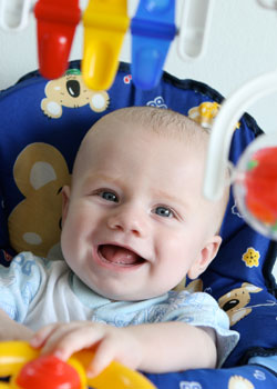 When your baby is about six months, you should take him to your doctor of optometry for his first thorough eye examination. Things that the optometrist will test for include excessive or unequal amounts of nearsightedness, farsightedness, astigmatism, lack of eye movement ability, as well as other eye health problems. These problems are not common, but it is important to identify children who have them at this stage. Vision development and eye health problems can be more easily corrected if treatment is begun early.
Unless you notice a need, or your doctor of optometry advises you otherwise, your child’s next eye exam should be around age three, and then again before he or she enters school.
During the first four months of life, your baby should begin to follow moving objects with the eyes and to reach for things, first by chance and later more accurately, as hand-eye coordination and depth perception begin to develop.
To help, use a nightlight or other dim lamp in your baby’s room; change the crib’s position frequently and your child’s position in it; keep reach-and-touch toys within your baby’s focus, about eight to twelve inches from his eyes; talk to your baby as you walk around the room; alternate right and left sides with each feeding; and hang a mobile above and outside the crib.
Between four and eight months, your baby should begin to turn from side to side and use his or her arms and legs. Eye movement and eye/body coordination skills should develop further and both eyes should focus equally.
Enable your baby to explore different shapes and textures with his or her fingers; give your baby the freedom to crawl and explore; hang objects across the crib; and play “patty cake” and “peek-a-boo” with your baby.
From eight to twelve months, your baby should become mobile, crawling and pulling himself or herself up. He or she will begin to use both eyes together to judge distances and grasp and throw objects with greater precision. To support development do not encourage early walking – crawling is important in developing eye-hand-foot-body coordination; give your baby stacking and take-apart toys; and provide objects your baby can touch, hold and see at the same time.
From one to two years, your child’s eye-hand coordination and depth perception will continue to develop and he or she will begin to understand abstract terms. Things you can do are to encourage walking; to provide building blocks, simple puzzles and balls; and to provide opportunities to climb and explore indoors and out.
There are many other affectionate and loving ways in which you can aid your baby’s vision development. Use your creativity and imagination. Ask your doctor of optometry to suggest other specific activities.
Pre-School Vision
 During the infant and toddler years, your child has been developing many vision skills and has been learning how to see. In the preschool years, this process continues, as your child develops visually guided eye-hand-body coordination, fine motor skills, and the visual motor skills necessary to learn to read.
As a parent, you should watch for signs that may indicate a vision development problem, including a short attention span for the child's age; difficulty with eye-hand-body coordination in ball play and bike riding; avoidance of coloring and puzzles and other detailed activities.
There are everyday things that you can do at home to help your preschooler’s vision develop as it should.
These activities include reading aloud to your child and letting him or her see what you are reading; providing a chalkboard, finger paints and different shaped blocks and showing your child how to use them in imaginative play; providing safe opportunities to use playground equipment such as a jungle gym and balance beam; and allowing time for interacting with other children and for playing independently.
There are everyday things that you can do at home to help your preschooler's vision develop as it should. These activities include:
These activities include reading aloud to your child and letting him or her see what you are reading; providing a chalkboard, finger paints and different shaped blocks and showing your child how to use them in imaginative play; providing safe opportunities to use playground equipment such as a jungle gym and balance beam; and allowing time for interacting with other children and for playing independently.
By age three, your child should have a thorough optometric eye examination to make sure your preschooler’s vision is developing properly and there is no evidence of eye disease. If needed, your doctor can prescribe treatment including glasses and/or vision therapy to correct a vision development problem.
Here are several tips to make your child's optometric examination a positive experience:
- Make an appointment early in the day. Allow about one hour.
- Talk about the examination in advance and encourage your child's questions.
- Explain the examination in your child's terms, comparing the E chart to a puzzle and the instruments to tiny flashlights and a kaleidoscope.
Unless recommended otherwise, your child's next eye examination should be at age five. By comparing test results of the two examinations, your optometrist can tell how well your child's vision is developing for the next major step. . . the school years.
School-Age Vision
 A good education for your child means good schools, good teachers and good vision. Your child's eyes are constantly in use in the classroom and at play. So when his or her vision is not functioning properly, learning and participation in recreational activities will suffer.
The basic vision skills needed for school use are:
- Near Vision: The ability to see clearly and comfortably at 10-13 inches.
- Distance Vision: The ability to see clearly and comfortably beyond arm's reach.
- Binocular Coordination: The ability to use both eyes together.
- Eye Movement Skills: The ability to aim the eyes accurately, move them smoothly across a page and shift them quickly and accurately from one object to another.
- Focusing Skills: The ability to keep both eyes accurately focused at the proper distance to see clearly and the ability to change focus quickly.
- Peripheral Awareness: The ability to be aware of things located to the side while looking straight ahead.
- Eye/Hand Coordination: The ability to use the eyes and hands together.
If any of these or other vision skills is lacking or does not function properly, your child will have to work harder. This can lead to headaches, fatigue, and other eyestrain problems. As a parent, be alert for symptoms that may indicate your child has a vision or visual processing problem. Be sure to tell your optometrist if your child frequently:
- Loses his place while reading
- Avoids close work
- Holds reading material closer than normal
- Tends to rub his eyes
- Has headaches
- Turns or tilts head to use one eye only
- Makes frequent reversals when reading or writing
- Uses finger to maintain place when reading
- Omits or confuses small words when reading
- Consistently performs below potential
Since vision changes can occur without you or your child noticing them, your child should visit the optometrist at least every two years, or more frequently if specific problems or risk factors exist. If needed, the doctor can prescribe treatment including eyeglasses, contact lenses, or vision therapy. Remember, a school vision or pediatrician's screening is not a substitute for a thorough eye examination.
Protective Eyewear
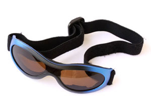 Please do not overlook the importance of safety eyewear when playing sports. Each year, hundreds of men, women, and children are injured when playing sports. To help prevent sports-related eye injuries, athletes should use protective athletic eyewear whether or not prescription eyewear is needed. One choice is a sports frame with prescription or non-prescription polycarbonate lenses. Baseball or softball players who are hit in or near the eye, or suffer a blow to the head, should seek immediate care at a hospital emergency room or from an eye care professional.
Children Contact Lenses
The important thing for parents and their children who wear contact lenses to remember is that contacts are prescribed medical devices. Contact lenses are not a cosmetic accessory. While the wearer may be happy about his or her new look, it is extremely important that the lenses be properly cleaned and worn according to the instructions of the optometrist.
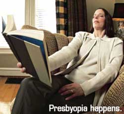
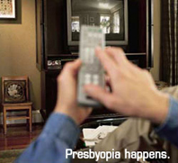 As people reach their mid to late 40s, a condition called presbyopia can set in. Presbyopia is the inability to focus on objects near the eye. One usually notices that it is harder to read or use the computer. Wearing bifocals or reading glasses is a way to remedy this condition.
Presbyopia is a natural consequence of the aging process. There is no cure, though researchers are constantly looking for one. Even if a person has never had vision problems before, he or she can still develop presbyopia. While symptoms can present suddenly, presbyopia usually occurs over a long period of time. Symptoms include having to hold things at arm’s length to see them clearly, eye strain, fatigue and headaches from near work.
Computer Glasses – To reduce eye strain and fatigue, we carry specialized computer lenses. These lenses are perfect for computer users who spend a majority of their days working on computers. And since three out of four computer users will suffer from Computer Vision Syndrome, computer lenses are a great way to keep your eyesight healthy.
Reading Glasses – One of the first areas of your life where presbyopia becomes prominent is in your ability to read. There are a variety of styles available, with sleek designs that allow you to carry them anywhere.
 No-Line Bifocals – For many presbyopes, bifocal lenses are a necessity. But it can be difficult to adjust to the harsh line that is found in bifocal lenses. Fortunately, there are no-line lenses, which are also called progressive lenses. No more lines! Just a gradual change in focusing power which allows you to comfortably focus on any distance. Just as in wearing bifocals, distant objects are viewed through the top portion of the lenses, and near objects are viewed through the bottom portion of the lenses.
Bifocal Contacts – If you need bifocals but cannot stand wearing glasses, you may need bifocal contact lenses. Now you can have all of the benefits of bifocal lenses in the convenience of contact lenses. Talk with your doctor about bifocal contacts today.
Monovision Correction – For some of our emerging presbyopes we offer another option to glasses, monovision. This is a method of fitting your dominant eye for distance vision and your non-dominant eye for near vision. Contacts are available in disposable, extended wear, and even daily disposable lenses to fit your lifestyle. Most patients require 2-4 weeks to make the adjustment from binocular vision to monovision.
Our top priority is the care of your eyes. We want to keep your eyes healthy through regular eye health evaluations, communication, and education. This page lists a few of the most common eye diseases. Select from the following list of topics or scroll to learn about the causes, symptoms, and treatments for:
| Blepharitis | Diabetic Retinopathy | Glaucoma |
| Cataracts | Dry Eye Syndrome | Macular Degeneration |
| Conjunctivitis | Retinal Detachment |
Blepharitis
 There are two types of blepharitis. Seborrheic blepharitis is often part of an overall skin condition called seborrhea, which may also affect the scalp, chest, back and the area behind the ears. The second form of blepharitis – staph blepharitis – is a more severe condition, caused by bacteria, that begins in childhood and may continue through adulthood.
Causes
Hormones, nutrition, general physical condition, and even stress may contribute to seborrheic blepharitis. Build-ups of naturally occurring bacteria contribute to staph blepharitis.
Symptoms
Blepharitis could be described as dandruff of the eyelids. Seborrheic blepharitis results in redness of the eyelids, flaking and scaling of eyelashes, and greasy, waxy scales caused by abnormal tear production. Staph blepharitis can cause small ulcers, loss of eyelashes, eyelid scarring, and even red eye.
Treatment
Careful cleaning of the eyelids can reduce seborrheic blepharitis. Application of hot packs to the eyes for 20 minutes a day can also help. Staph blepharitis may require antibiotic drops and ointments.
Cataracts
 A cataract is a cloudiness that occurs in the lens of the eye. The lens is made mostly of water and protein that is arranged to let light through. Sometimes the protein clumps, blocking light and making the lens appear cloudy.
Symptoms
A person with cataracts may encounter faded colors, problems with light (such as halos, or headlights that seem too bright), poor night vision, double vision, or multiple vision.
Treatment
Your eye doctor can detect the presence of cataracts through a thorough eye exam, including a visual acuity test and dilation of the pupils. Treatment is available to prevent or reduce cataracts.
Conjunctivitis
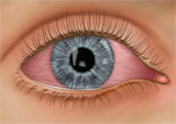 Conjunctivitis, commonly called pink eye, is a redness of the eye. It is often accompanied by a discharge (clear, yellow, or white) and itching in the eye.
Causes
Pink eye is most often a viral infection, but it may also be caused by bacteria or an allergic reaction. Viral pink eye is highly contagious.
Prevention and Treatment
To avoid spreading conjunctivitis, wash your hands often, do not touch the infected area with your hands, do not share wash cloths or towels, and avoid using makeup which may become contaminated. A child with pink eye should be kept from school for a few days. Sometimes an eye doctor will need to prescribe antibiotic eye drops and ointments to clear up conjunctivitis.
Diabetic Retinopathy
Diabetic retinopathy is a condition associated with diabetes. High levels of blood sugar may damage tiny blood vessels in your eye. New vessels may form to replace the damaged vessels. The new vessels can burst, resulting in blurred vision or even blindness.
Symptoms
Symptoms of diabetic retinopathy include:
- "Floaters” – small specks that pass across your field of vision, made up of cells floating in the transparent gel of your eyeball
- Difficulty reading or seeing things close-up
- Sudden loss of vision
- Flashes
- Blurred or darkened vision
Risk Factors and Treatment
If you have diabetes, make sure you control your blood sugar level. This will reduce your risk of getting diabetic retinopathy. If you are experiencing some of the symptoms listed above, give us a call. If diagnosed properly, diabetic retinopathy can be treated with a laser procedure or a vitrectomy.
Dry Eye Syndrome
 If your eyes are constantly itchy or dry, you may have dry eye syndrome, which affects almost 10 million Americans. Dry eye syndrome is caused by a lack of, or poor quality of, tears. Tears lubricate the outer layer of the eye called the cornea. If the tears are not composed of a proper balance of mucous, water, and oil, the eye becomes irritated.
Symptoms
Dry eye syndrome leads to a number of symptoms, including itchiness, irritation, burning, excessive tearing, redness, blurred vision that improves with blinking, and discomfort after long periods of watching television, using a computer, or reading.
Risk Factors
There are many factors that can contribute to dry eye syndrome. These include dry, hot, or windy climates; high altitudes; air-conditioned rooms; and cigarette smoke. Contact lens wearers, people with abnormally dry skin, and the elderly are more likely to develop dry eye syndrome. You may also be more at risk if you take certain medications, have a thyroid condition, a vitamin-A deficiency, Parkinson’s or Sjorgen’s disease, or if you are a woman going through menopause.
Glaucoma
 Glaucoma is a very common eye disorder affecting millions of Americans. It is caused by too much pressure on the inside of the eye. The fluid in your eyes helps to nourish and cleanse the inside of your eyes by constantly flowing in and out. When the fluid is prevented from flowing out, the intraocular pressure builds and damages the optic nerve. This causes a gradual loss in peripheral vision.
Symptoms
Those suffering from open-angle glaucoma experience a type of tunnel vision, where their field of vision gradually decreases. It can eventually lead to blindness. Narrow-angle glaucoma, which is rare, carries symptoms of sharp pain in the eyes, blurred vision, dilated pupils, and even nausea or vomiting. It can cause blindness in a matter of days, and it requires immediate medical attention.
Risk Factors
Heredity seems to be a risk factor. Also, you may be at greater risk if you are over 45, of African descent, near-sighted, or diabetic. Finally, if you have used steroids or cortizone for a long period of time, or if you have suffered an eye injury in the past, you have a greater chance of developing glaucoma.
Macular Degeneration
Macular degeneration is a disease which affects a small area of the retina known as the macula. The macula is a specialized spot on the retina that allows us to see the fine detail of whatever is directly in front of us. Macular degeneration occurs when the macula begins to deteriorate.
“Wet” vs. “Dry”
Most often, macular degeneration is accompanied by formation of yellow deposits, called “drusen,” under the macula, which dry out or thin the macula. This is called “dry” macular degeneration. In rare cases, abnormal blood vessels develop under the macula and leak fluid. This is called “wet” macular degeneration.
Causes
A number of uncontrollable factors contribute to macular degeneration, including age, sex, eye color, farsightedness, and race. Risk factors you can control include smoking, high blood pressure, exposure to harmful sunlight, and diet.
Symptoms
It is difficult to detect dry macular degeneration in its early stages. The most common symptoms, when detected, include a spot of blurry vision, dark vision, or distorted vision. Wet macular degeneration progresses much faster than the dry variety. Both forms of macular degeneration can cause blindness.
Treatment
Currently, there is no cure for macular degeneration, but treatment is available to slow the effects.
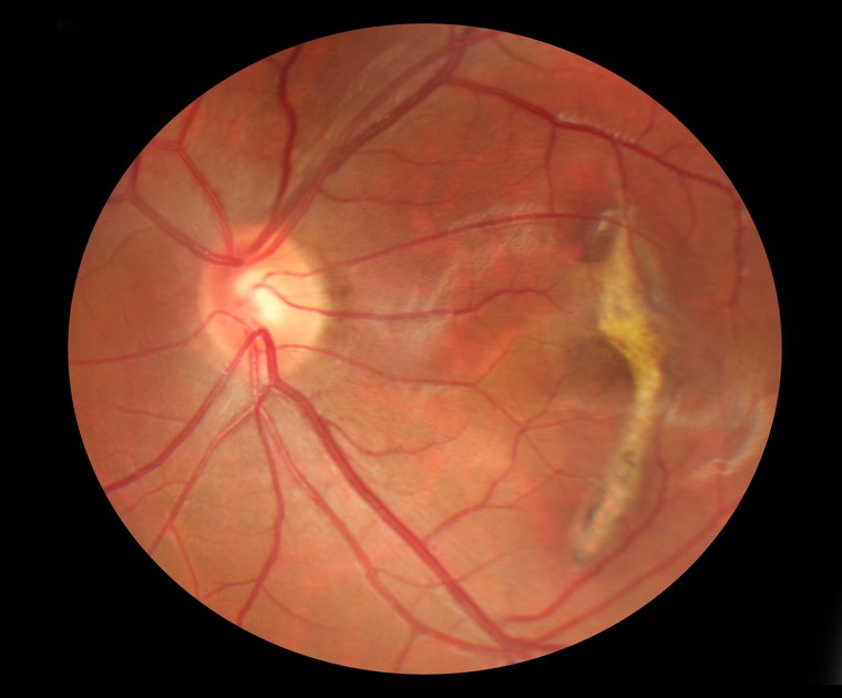
Retinal Detachment
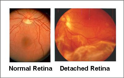 The part of the eye which collects light and transmits the light messages to the optic nerve and brain is the retina. It lines the inner back wall of the eye. When the retina separates from the back wall, it is known as retinal detachment. It is a serious condition which can cause permanent damage and vision loss if not treated quickly.
Symptoms
A retinal detachment often causes sudden defects in your vision. It may just cause a blind spot too small to notice, or it may cause a noticeable shadow which obscures your vision. An increase in “floaters,” which look like small particles or fine threads, may also be noticed. Finally, flashes of light are associated with retinal detachment.
Risk Factors
Eye injuries, tumors, and cataract surgery can cause retinal detachment. Near-sighted individuals and the elderly are at greater risk for spontaneous detachment. Also, diabetic retinopathy, a condition associated with diabetes, can cause bleeding which leads to retinal detachment.
We are happy to provide you with some basic information on several common vision conditions. Select from the following list or scroll to learn more about the symptoms and treatments for:
| Hyperopia | Emmetropia |
| Astigmatism | Myopia |
| Computer Vision Syndrome | Presbyopia |
Amblyopia (lazy eye)
 Focus on Amblyopia
Amblyopia, commonly called lazy eye, occurs when one eye develops differently than the other eye does, causing one eye to be weaker than the other. Sometimes a difference in focusing ability causes one eye to be used more often. Other times, the eyes are misaligned, causing one eye to “shut off” to avoid double vision. Regardless of the cause, the result is a weakened, or amblyopic, eye.
Focus on Symptoms
It’s hard to spot amblyopia. Sometimes a child will noticeably favor one eye over the other. Another possible symptom is the child frequently bumping into things on one side. The best way to tell if your child has lazy eye is through a complete exam at about six months and three years. Early diagnosis can prevent amblyopia from leading to more serious problems, such as loss of the ability to see three dimensions or functional blindness in the amblyopic eye.
Focus on Treatment
Most of the time amblyopia cannot be entirely corrected. The amblyopic eye will always be a bit weaker than the other. However, with treatment, vision in the amblyopic eye can be improved to some extent. Treatment involves encouraging the weak eye to develop. This is done using eye patches, vision therapy, glasses, or a combination of the three. The strong eye may be patched to encourage the weak eye to develop. Vision therapy can help to correct improper use of the eyes. If a focusing error is at the root of the problem, then glasses may reduce the error. Most of the time the amblyopic eye will always require glasses.
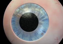
Astigmatism
 Sometimes the cornea is irregularly shaped, causing the eye to focus an object on two different areas of the retina. This is known as astigmatism. For the cornea to bend light correctly, it should be dome-shaped, like a basketball. Astigmatic corneas are shaped more like a football. This causes a distorted view when looking at objects which are close-up and far away.
The cause of astigmatism is unknown. Astigmatism is often associated with myopia or hyperopia, and it usually is present from birth. It may be hereditary, or it may be caused by factors such as pressure on the cornea, incorrect posture, or increased use of the eyes for “near work.” Mild astigmatism usually does not need to be corrected. Eyeglasses, contact lenses, or refractive surgery can correct moderate to high degrees of astigmatism.
Presbyopia - Eyes Over 40
 As a people get older, usually when they hit their mid to late 40s, a condition called presbyopia can set in. Presbyopia is the inability to focus on objects near the eye. One usually notices that it is harder to read or use the computer. Bifocals or reading glasses are a way to remedy this condition.
Presbyopia is a natural consequence of the aging process. There is no cure, though researchers are constantly looking for one. Even if a someone has never had vision problems before, he can still develop presbyopia. It may seem to occur suddenly, but it actually occurs over a long period of time. Symptoms include having to hold things at arm’s length to see them clearly, eye strain, fatigue, and headaches from near work.
Fortunately, we carry a number of products designed to eliminate the difficulties associated with presbyopia.
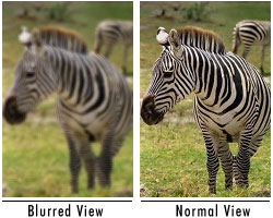
Computer Vision Syndrome
 Focus on Computer Vision Syndrome
Computer vision syndrome (CVS) affects three out of four computer users. It is a series of symptoms related to extended periods of computer usage. Though it is no cause for panic, measures can be taken to relieve symptoms of CVS.
Focus on Symptoms
CVS can appear as a variety of symptoms. Headaches, eye strain, neck and back aches, sensitivity to light, blurred vision, double vision, and dry or irritated eyes are all possible problems related to CVS.
Focus on Risk Factors
Any computer user can develop CVS. Your vision, your computer, and the environment where you use your computer are all factors which can lead to CVS.
Computer Glasses - To reduce eye strain and fatigue, we carry specialized computer lenses. These lenses are perfect for computer users who spend a majority of their day working on a computer. And since three out of four computer users will suffer from Computer Vision Syndrome, computer lenses are a great way to keep your eyesight healthy.
Emmetropia
When rays are focused correctly on the retina of a relaxed eye, the eye is said to be emmetropic. Emmetropia is the medical term for 20/20 vision, vision that needs no corrective lenses, contact lenses, or reading glasses. It occurs because the optical power of the eye can perfectly focus an image to the retina, giving it “perfect” vision.
The opposite of emmetropia is ametropia. With ametropia, the focal point of the eye is some distance in front of or behind the retina. The following vision conditions are types of ametropia.
Hyperopia (farsightedness)
 Hyperopia is more commonly known as farsightedness. As the name suggests, people with farsightedness are able to focus on objects that are further away, but have difficulty focusing on objects which are very close. This is because the eyeball is shorter than normal, which prevents the crystalline lens in the eye from focusing correctly on the retina. About a fourth of the population is farsighted. Hyperopia can lead to chronic glaucoma, a more serious condition, later in life.
A family history of hyperopia is a risk factor for developing hyperopia. Babies are often born with hyperopia but they can usually outgrow the condition as their eyes develop into the correct shape.
Hyperopia can be corrected with eyeglasses or contact lenses. There are also new surgical procedures that can correct hyperopia.
Myopia (nearsightedness)
 Myopia is the medical term for what most people call nearsightedness. It is a condition in which a person can see objects clearly only when they are close; when objects are farther away it is difficult to focus on them. Myopia usually develops in early childhood, although it sometimes develops in early adulthood. In rare cases, myopia can lead to more serious conditions such as retinal detachment.
Myopia is considered a genetic disorder. If your parents are nearsighted, you are at greater risk of also being nearsighted. Another risk factor is “near work” – work involving fine detail or focusing on close objects.
Myopia can be accommodated and sometimes corrected with eyeglasses or contact lenses. Sometimes myopia continues to gradually worsen throughout life, a condition known as myopic creep. Myopia can also be corrected by LASIK surgery.
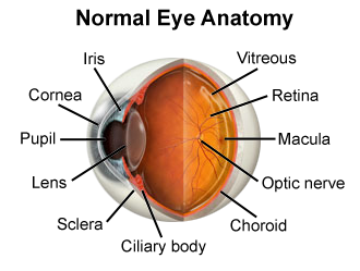
Don’t remember the lessons on eye anatomy from your highschool biology class? That’s OK—we have provided the following eyeball illustration and terms just to give you a refresher course. And we won’t give you a pop quiz afterwards…
| IRIS: | Pigmented tissue lying behind cornea that (1) gives color to the eye, and 2) controls amount of light entering the eye by varying size of black pupillary opening; separates the anterior chamber from the posterior chamber. |
| CORNEA: | Transparent front segment of the eye that covers iris, pupil, and anterior chamber, and provides most of an eye's optical power. |
| PUPIL: | Variable-sized, circular opening in center of iris; it appears as a black circle and it regulates the amount of light that enters the eye. |
| LENS: | Natural lens of eye; transparent intraocular tissue that helps bring rays of light to focus on the retina. |
| SCLERA: | The white of the eye; a protective fibrous outer layer covers all of the eyeball except for the part covered by the cornea. |
| CILIARY BODY: | A muscular ring under the surface of the eyeball; helps the eye focus by changing the len’s shape and also produces aqueous humor. |
| CHOROID: | The vascular layer between the sclera and the retina; the blood vessels in the choroid help provide oxygen and nutrients to the eye. |
| OPTIC NERVE: | Largest sensory nerve of the eye; carries impulses for sight from retina to brain. |
| MACULA: | Small, specialized central area of the retina responsible for acute central vision. |
| RETINA: | Part of the eye that converts images into electrical impulses sent along the optic nerve for transmission back to the brain. Consists of many named layers that include rods and cones. |
| VITREOUS: | Transparent, colorless, gelatinous mass; fills rear two-thirds of the interior of the eyeball, between the lens and the retina. |